The uterus is composed of three layers — the perimetrium, myometrium, and endometrium — each fulfilling specific functions.
The perimetrium (serosal layer) forms the outer portion of the uterus and secretes fluid to reduce friction with nearby organs. The myometrium (muscular layer) makes up most of the uterine volume and consists of smooth muscle cells. It is thick and firm, expanding during pregnancy to accommodate the growing fetus and contracting during childbirth to enable delivery. The endometrium (mucosal layer) is the inner lining of the uterus and undergoes cyclical changes throughout a woman’s life.
During the menstrual cycle, under hormonal influence, the endometrium thickens to receive the blastocyst (fertilized egg), which implants into this layer. During pregnancy, the glands and blood vessels within the endometrium continue to grow, and vascular spaces merge to form the placenta, which supplies oxygen and nutrients to the embryo and, later, the fetus. In the absence of fertilization, the superficial layer of the endometrium sheds and is eliminated as menstrual bleeding. At menopause, the endometrium becomes atrophic.
The appearance and thickness of the endometrium depend largely on reproductive age and the phase of the menstrual cycle. Normally, the endometrium measures around 6–7 mm, gradually thickening and reaching its maximum just before menstruation.
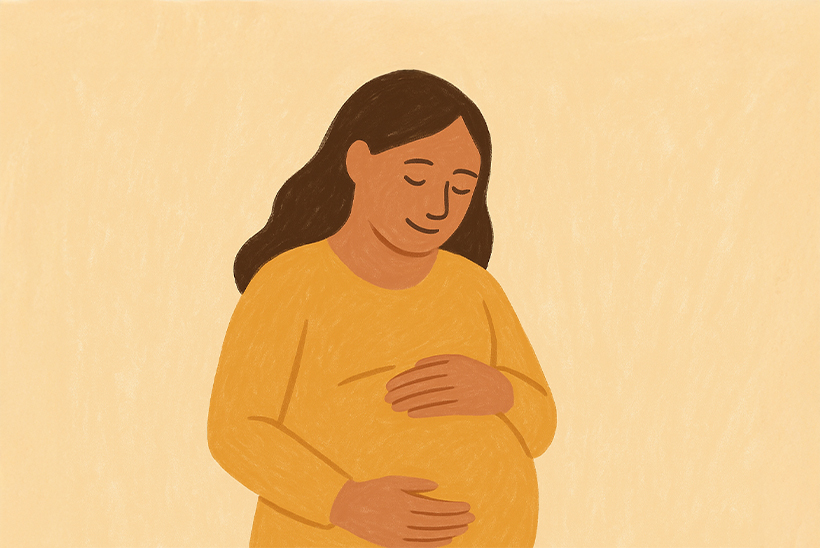
“You deserve to be heard, seen, treated with respect, and supported throughout your life.”

Women with consistently thickened endometrium (endometrial hyperplasia) often face difficulties conceiving. This condition requires medical evaluation to determine the underlying cause. The most frequent explanation is excessive estrogen combined with inadequate progesterone. Hormone therapies may also cause abnormal thickening. Contributing factors include overweight and obesity due to excessive estrogen production. Occasionally, an estrogen-secreting ovarian tumor may lead to an abnormally thick endometrium.
A thin endometrium can also contribute to infertility. Studies show that temporary thinning of the endometrium may occur after long-term use of oral contraceptives.
Normal endometrial thickness values on transvaginal ultrasound are:
- During menstruation: 2–4 mm
- Early proliferative phase: 5–7 mm
- Late proliferative preovulatory phase: up to 11 mm
- Secretory phase: 7–16 mm
- After dilation, curettage, or miscarriage: under 5 mm
Talk with me about
Endometrial Conditions

Similar Articles

In Vitro Fertilisation (IVF) | The Patient Experience
Five Things You Should NOT Do After Embryo Transfer